
Hemangioma of the Buccal Mucosa of the Cheek Presenting as a Dermoid Cyst: A Case Report
Abstract
This case report presents a 73-year old Asian female with a presumed dermoid cyst in the buccal mucosa of the left cheek that was visible and embarrassing to her. Histopathology revealed the mass to be consistent with a cavernous hemangioma. Hemangiomas are benign vascular tumors of mesenchymal origin. It is usually present at birth but can develop later in life. It commonly occurs in the head and neck region, but rarely in the oral cavity.
Author Contributions
Academic Editor: Qiping Dong, China
Checked for plagiarism: Yes
Review by: Single-blind
Copyright © 2023 Cameron Y. S. Lee
 This is an open-access article distributed under the terms of the Creative Commons Attribution License, which permits unrestricted use, distribution, and reproduction in any medium, provided the original author and source are credited.
Competing interests
The authors have declared that no competing interests exist.
Citation:
Introduction
Hemangiomas are a group of benign mesenchymal vascular tumors characterized by a proliferation of blood vessels histologically.1Based on histologic examination, hemangiomas are classified as either capillary or cavernous benign vascular tumors. Hemangiomas are commonly observed soft tissue tumors in the head and neck region.2, 3In the oral cavity, hemangiomas are rarely observed. Clinically, hemangiomas may appear as a painless submucosal papule or nodule single or lobulated mass that is bluish-purple in color.4We report a case of a 73-year old Asian female who presented with a submucosal mass in the left buccal mucosa of the left cheek that clinically appeared to be a dermoid cyst. However, during surgery to excise the lesion the dome-shaped mass observed was a dark purple vascular tumor. Histopathologic examination confirmed the lesion as a cavernous hemangioma.
Case Report
A 73-year old Asian female presented to the office for evaluation of a mass on the left cheek (Figure 1). Medical history reported by the patient included hypertension and osteoporosis. Medications included Losartan 50mg daily and Alendronate 70mg once per week. The patient stated that the mass appeared to be enlarging in size and was noticeable to her family. She denied pain or tenderness in the area of the cheek mass. Oral examination revealed a firm compressible submucosal mass in the left buccal vestibule of the mandible that did not blanche on digital pressure. The mass was negative for any bruit or vascular thrill. The surface mucosa overlying the mass was the same color as the buccal mucosa of the cheek (Figure 2). No imaging studies were indicated, as the mass was localized to the soft tissues of the cheek. The differential diagnosis included dermoid cyst, epidermoid cyst, and mucous retention cyst. The patient was informed of the clinical findings and the plan to remove the lesion as an in-office procedure. Under local anesthesia, using a #15 scalpel a mucosal incision was made and it was immediately observed that the dark purple mass was a suspected hemangioma (Figure 3). With careful blunt dissection, the purple-colored mass was completely excised and sent for histologic analysis (Figure 4). Histopathologic examination revealed a proliferation of irregular dilated capillary sinuses lined by flat endothelial cells. The sinusoidal spaces contained erythrocytes (Figure 5). The diagnosis was consistent with a cavernous hemangioma.
Figure 1.Clinical photograph of subtle mass on left cheek (arrows) that appeared to be a dermoid cyst.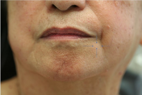
Figure 2.Intraoral photograph of mass in left buccal mucosa of cheek. Note the normal pink color overlying the mass.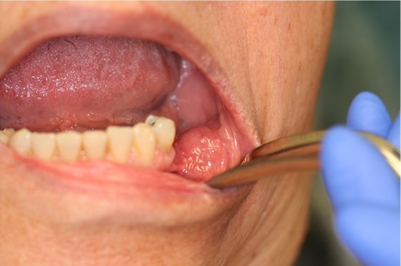
Figure 3.Intraoperative photograph of dissection of hemangioma from left cheek.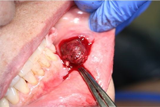
Figure 4.Completely excised surgical specimen.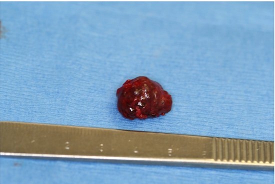
Figure 5.Histopathology of cavernous hemangioma. Note irregular sinusoids lined by endothelium containing erythrocytes. Hematoxylin and eosin, 400x.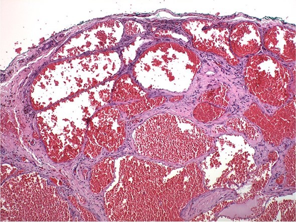
Discussion
In the oral cavity, cavernous hemangiomas are rare painless dark purple-blue colored soft tissue masses that will blanch with digital pressure. Hemangiomas can occur on the gingiva, soft and hard palate of the maxilla, lips, buccal mucosa, salivary glands, and jaw bones.4, 5They can appear as a smooth single or multiple lobulated compressible mass of variable size.1, 6, 7However, hemangiomas can mimic other tumor or cystic lesions as in this case report. In our patient, the surface mucosa was of normal color without the dark purple-blue color that is characteristic of hemangiomas. Histopathology reveals well developed capillary sinuses lined by endothelial cells separated by a connective tissue stroma. The dilated sinuses are often filled with erythrocytes.4, 8
Management of hemangiomas are not always indicated. When treatment is initiated, age of the patient, size and extent of the lesion must be considered.4Treatment options include surgical excision, intralesional injection of sclerosing agents, laser surgery, cryosurgery, and interferon treatment.9, 10, 11, 12
Conclusion
Although rarely observed in the oral cavity, this case report describes a hemangioma that presented as a dermoid cyst in the buccal mucosa of the cheek. Intraoperatively, it was determined that the mass was a hemangioma. Histopathology confirmed that the lesion was a cavernous hemangioma.
References
- 1.Dilsiz A, Aydin T, Gursan N. (2009) Capillary hemangioma as a rare benign tumor of the oral cavity: a case report. Cases J. 2(1), 8622.
- 2.Warner M, Suen J Y. (1999) A classification of congenital vascular lesions. Hemangiomas and vascular malformations of the head and neck In: Warner M, Suen JY, editors , San Francisco; Wiley; 1-12.
- 3.Donald P J. (2001) Vascular anomalies of the head and neck. Facial Plast Surg Clin North Am. 9, 77-92.
- 4.Neville B W, Damm D D, Allen C M. (2002) Oral and Maxillofacial Pathology. , Philadelphia: 447-449.
- 5.Bonet-Coloma C, Minguez-Martinez I, Palma-Carrio C. (2011) Clinical characteristics, treatment and outcome of 28 hemangiomas in pediatric patients. Med Oral Pathol Oral Cir Bucal. 16(1), 19-22.
- 6.Delaney J E, Keels M A. (2000) Pediatric oral pathology: soft tissue and periodontal conditions. , Pediatric Clin N Am 47(5), 1125-1147.
- 7.VRV Kumari, Vallabhan C G, Geetha S. (2015) Atypical presentation of capillary hemangioma in oral cavity. A case report. , J Clin Diagn Res 9(10), 26-8.
- 8.Kamala K A, Ashok L, Sujatha G P. (2014) Cavernous hemangioma of the tongue: a rare case report. Contemp Clin Dent. 5(1), 95-98.
- 9.Chin D C. (1983) Treatment of maxillary hemangioma with sclerosing agent. Oral Surg Oral Med Oral Pathol. 55, 247-249.
- 10.Barak S, Katz J, Kaplan I. (1991) The CO2laser in surgery of vascular tumors of the oral cavity in children. , J Dent Child 58, 293-296.